
To contact: David Legrady, Csilla Pesznyak
Radotherapy
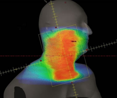 Radiotherapy plays a key role in treating tumor patients. The two main types of radiotherapy are teletherapy (external radiotherapy, application of MeV-energy ionizing radiation) and brachytherapy (internal radiotherapy, insertion of radioactive isotopes into the body). Radiation damages the reproduction information (DNA) of tissues, therefore replication of cancer cells decrease. Biological changes caused by radiation is described by radiation biology models accounting for both the regeneration of healthy tissues and the elimination of cancer cells. Radiotherapy treatment targets the most efficient eradication of cancer cells while ensuring the protection of healthy tissues. Accordingly, different treatment planning methods may be used for the best delivered dose distribution.
Apart from theoretical education, practical courses are organized at the National Institute of Oncology , and students can participate in research work. The students learn how to use radiotherapy treatment planning systems, further specialized techniques like intensity modulated, image guided radiotherapy and stereotactic radiosurgery. Measurement methodology applied to radiotherapy is very diverse: several types of ionization chambers, film dosimetry, semiconductor detectors, gel dosimetry and special phantoms are used, all available for student and research use. Before treatments, Quality Assurance dosimetry measurements must be performed, these measurement techniques are also accessible for the students.
Magnetic Resonance Imaging
Pulse Sequence Development for Siemens Human scanner
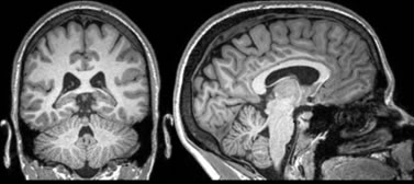 Method development is ongoing at the ELKH TTK Brain Imaging Centre using a Siemens 3T human MRI scanner. The multiband layout offers options for sequence development targeting improvement of the Signal-to-Noise ratio. Further, functional MRI and mapping special brain tissue types is also aimed at.
Pulse Sequence Development for Mediso preclinical PET-MRI scanner
Within the >>„VKSZ_14 - K+F Versenyképességi és Kiválósági Szerződések” VKSZ-14-0151<< project pulse sequence development started for a preclinical small animal scanner.
MRI hardware development for a Bruker NMR magnet
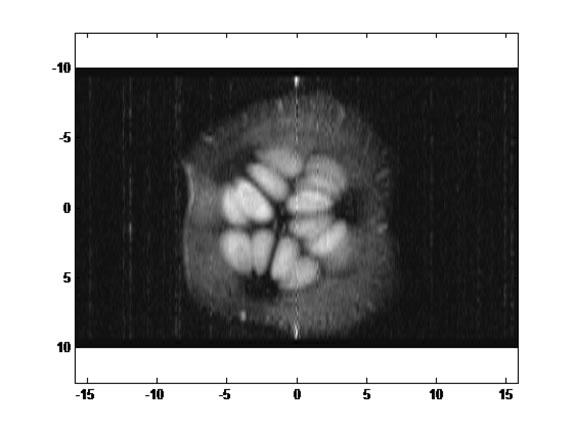
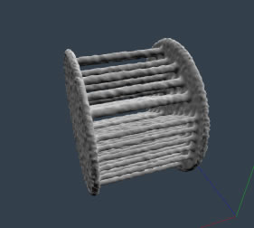
Together with the BME Institute of Physics a MRI machine was put together based on a Bruker Avance 7T NMR magnet. The equipment is capable of imaging object with volume of a few cm3. Illustration shows the MRI image of a slice of a cucumber taken with this machine.
A Magritec Terranova student demonstration equipment is also available for the students.
Altogether four MRI scanners are accessible for the students to experiment with the practical side of MRI from BSc level up to PhD research.
Nuclear Medicine
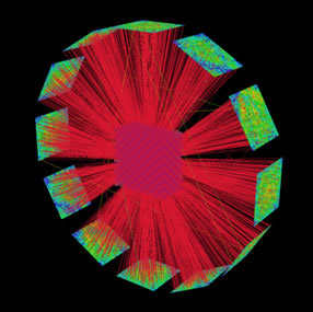 Within the Teratomo Project, an ML-EM based Positron Emission Tomography reconstruction code, PANNI (PET-Aimed Novel Nuclear Imager) development was started. The specialty of this code is that at each iteration step a full Monte Carlo simulation ensures that every aspect of PET imaging is accounted for.
 The computation power required for these simulations are provided by GPU’s, following the life of at least 108 particles /s. The Monte Carlo engine contains state of the art variance reduction techniques. Illustrations: trajectories in a PET-geometry; reconstructed image of a Derenzo-phantom.
Transmission Imaging: planar and Tomography
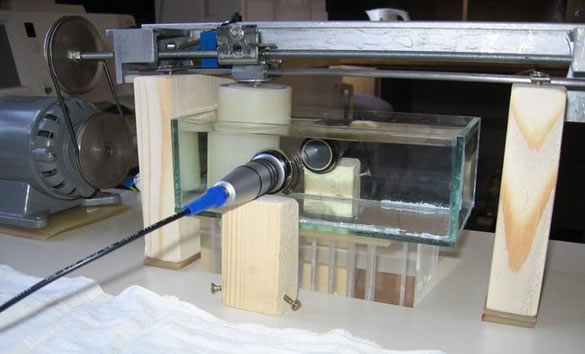
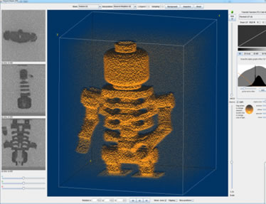 In the framework of „ KMOP-4.2.1/B-10-2011-0011” a Source-Ray 35-80kV, 80W x-ray source and a Dexela 1207 detector module was acquired and later built into a cone-beam micro CT scanner (NTIuCT) and for planar acquisition.
[illustrations: photo and x-ray planar image of a painting; measurement setup]
Ultrasound Diagnostics
 Ultrasnound Diagnostics is currently restricted to student practices and theoretical classes. Illustration: Sono-CT scanning at the institute.